
Mississippi State is a pioneer in using imaging technologies for veterinary patients. Earlier disease detection leads to improved treatment outcomes, which is great news for dogs and cats — and the people who love them.

Mississippi State has great news for pet owners: Many imaging techniques used to treat diseases in people also work on dogs and cats.
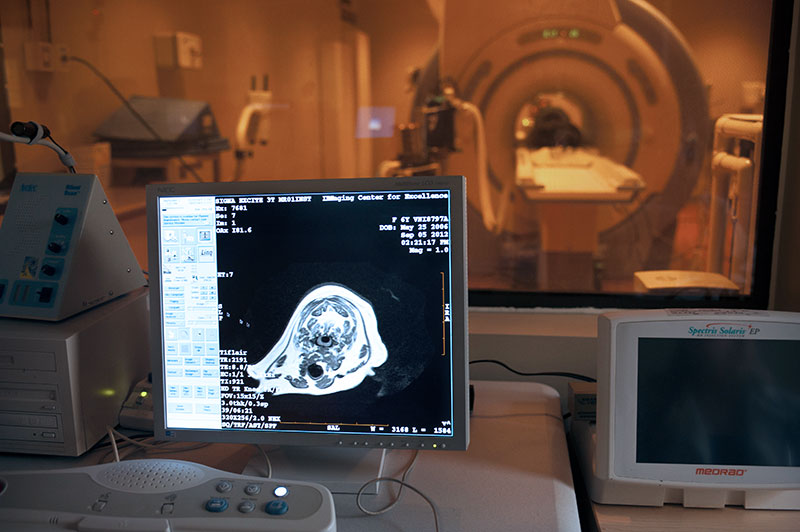
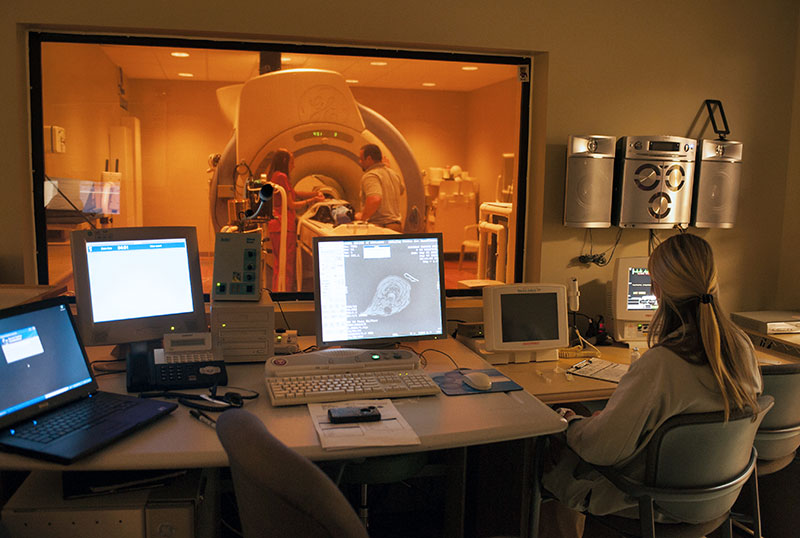
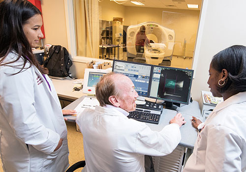
Most are familiar with MRIs and CT scans — common imaging tools that revolutionized early disease detection in humans. By adapting proven imaging technologies to help veterinary patients, MSU’s College of Veterinary Medicine continues making life-saving breakthroughs in the diagnosis and treatment of pet diseases such as cancer and epilepsy.
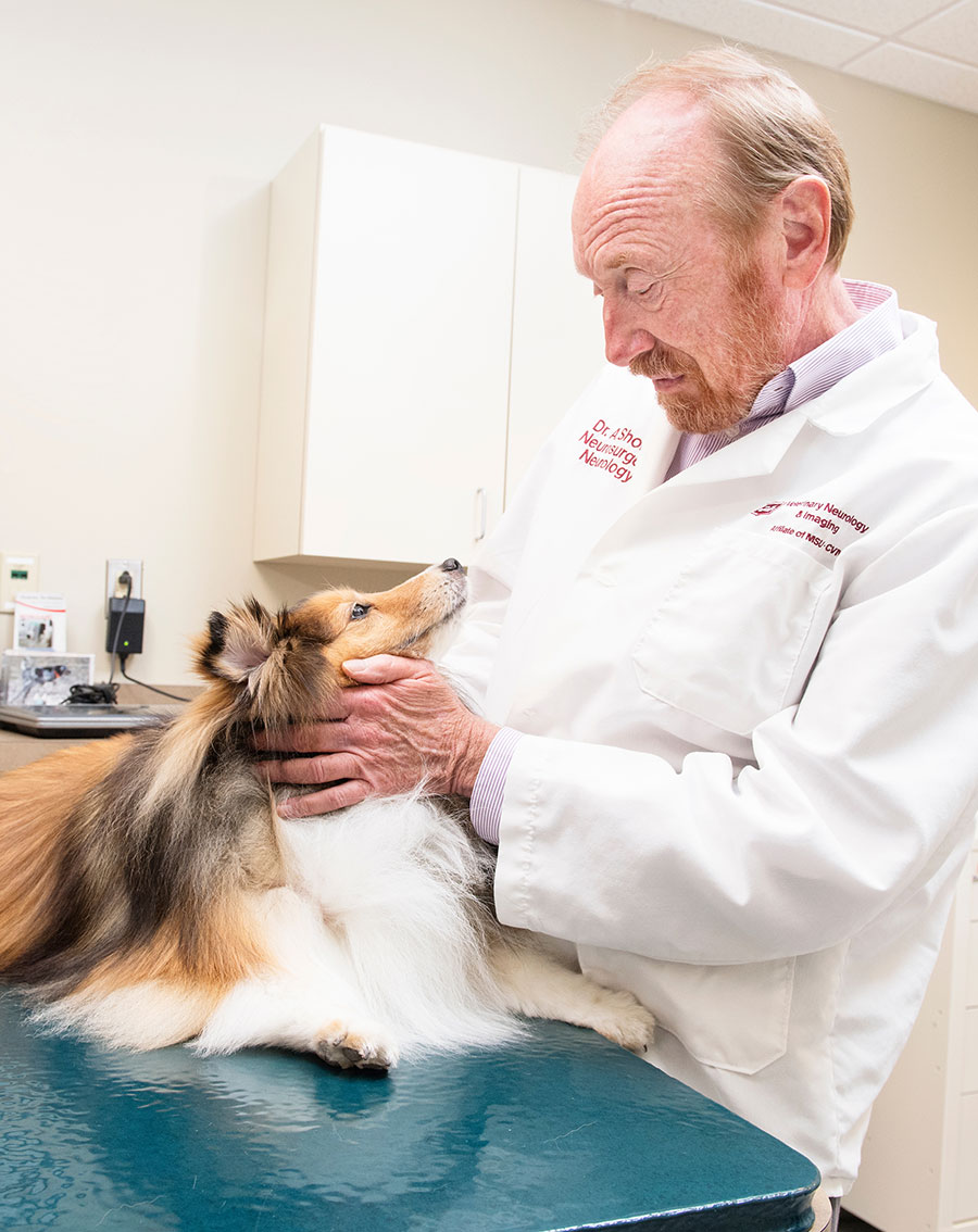
The Journal of the American Veterinary Medical Foundation has highlighted MSU’s use of state-of-the-art imaging technologies for neurology and oncology patients at the university’s Veterinary Specialty Center. Since the article’s publication, MSU veterinarians have made even more advancements in imaging technology to diagnose and treat pet diseases, says Dr. Andy Shores, clinical professor and service chief for neurosurgery and neurology.
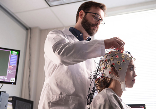
One advancement is magnetic resonance spectroscopy, or MRS.
“MRS is an imaging technique that evaluates the chemical and mineral content of tumor tissue,” explains Shores. “MRS allows us to isolate brain areas where we see a tumor or abnormal tissue and determine the type of tumor we’re dealing with so we can develop the best course of treatment.”
MRS also is effective for analyzing the brains of dogs with idiopathic epilepsy, having no known case or association with tumors, genetics or other seizure-inducing conditions. By solving the mysteries of idiopathic epilepsy, MSU veterinarians hope to improve treatment outcomes for dogs and cats that suffer from seizures and perhaps deepen understanding of the disease in humans.
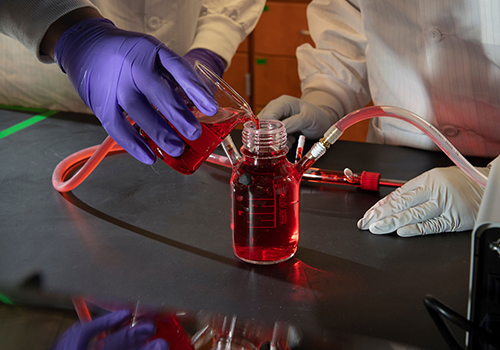
MSU’s veterinary research also holds exciting possibilities for breakthroughs in human cancer treatments. MSU is among a select group of veterinary schools testing a new treatment for brain tumors known as gliomas. Funded by a grant from the National Institutes of Health, the procedure involves injecting a modified virus into post-operative tumor sites in dogs. The virus is programmed to invade and eventually kill off lingering tumor cells in the brain while leaving normal cells unharmed.
After MSU veterinarians use imaging to diagnose glioma brain tumors, the NIH grant covers the cost of surgery and hospitalization for canine patients.
For prospective students interested in starting their veterinary careers at MSU, the variety of research projects coupled with access to the industry’s most advanced equipment and treatment techniques are strong recruiting tools. MSU’s learning environment also has been beneficial for faculty members interested in expanding their knowledge and experience.
“Mississippi State provides opportunities to do a lot of different things and to be a free thinker,” says Shores, who in 2017 co-edited the first textbook entirely devoted to the field of veterinary neurosurgery.